Brain Museum

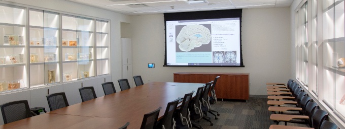
Interior of the Brain Museum on the ground floor of the Jacobs School building.
Visitors to these facilities located on South Campus and in the Jacobs School of Medicine and Biomedical Sciences get a learning experience that’s literally more in-depth than a traditional slide- and textbook-based education.


Christopher S. Cohan, PhD, curator of the Brain Museum, shows some specimens to interested medical students at the museum’s South Campus location in the Biomedical Education Building.
A World-Class Collection
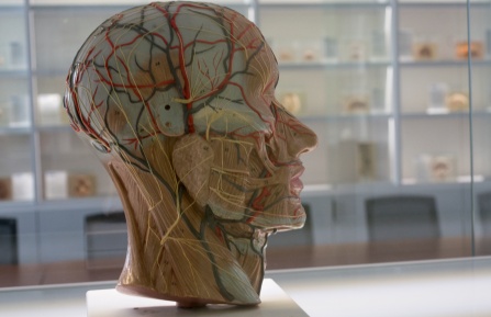
Taken as a whole, the Brain Museum’s collection demonstrates how the components of our nervous systems work together. Visitors can see the brain’s layers and internal structures firsthand and view them from different angles.
At the original South Campus location, almost 90 beautifully illuminated brain specimens highlight anatomical features such as the corpus callosum, hippocampus and cerebellum. Pathological specimens show conditions such as Alzheimer’s disease, cerebral aneurysms and hydrocephalus.
Dissections show the full pathways for vision and hearing, and photographs offer closer views of the brain’s intricate structures. A display that highlights the achievements of groundbreaking researchers in neuroanatomy sets the discipline in its historical context.
The museum also houses a world-class collection of slides that display stained cross-sections of brain tissue, which medical students and researchers can consult by arrangement with the Department of Pathology and Anatomical Sciences.

Founded in 1994, our Brain Museum is the only one in the United States dedicated exclusively to the brain.
Second Location Downtown Expands Resources
A second museum has been created with all new specimens in the Jacobs School’s building on the Buffalo Niagara Medical Campus. The downtown museum has about 40 human brain specimens, as well as various anatomical models of brains, skulls and other items. Digital displays have been installed at the front of the room that will eventually provide information about specimens in the museum and have the capability to be interactive and show 3D-images of brains.
This provides a convenient resource for our medical students while they are studying the brain. It also provides a resource for visitors to the Jacobs School building.
Some of the downtown brain specimens are modeled after the South Campus specimens, but others are unique. The downtown location, ability to use digital displays, and convenient location near the building entrance are important differences from the South Campus museum that will provide better accessibility and a different experience for visitors.
Neuroscience for the Community
The museum’s founder and long-time curator, Harold Brody, MD ’61, PhD, prepared many of the specimens and slides himself. A former professor and chair of anatomy and cell biology, Brody built this exhibit to be used by everyone, from kindergartners to neurosurgery students.
Today, Christopher S. Cohan, PhD, Distinguished Professor Emeritus, carries on Brody’s legacy. He made the museum a core resource for students in his neuroscience classes and conducts tours for interested groups, which have included our own MD and PhD students, dental and occupational therapy students, psychology PhD candidates, undergraduate students, guests at parents’ weekend and Girl Scouts.
Locations
955 Main Street, Room 1110
Buffalo, N.Y. 14214
360 Biomedical Education Building
University at Buffalo, South Campus
3435 Main Street
Buffalo, N.Y. 14214
Tours
To arrange a guided tour, contact:

