The Health Sciences Collaborative Core Facilities (HSCCF) provide multipurpose services to the biomedical research community at the University at Buffalo.
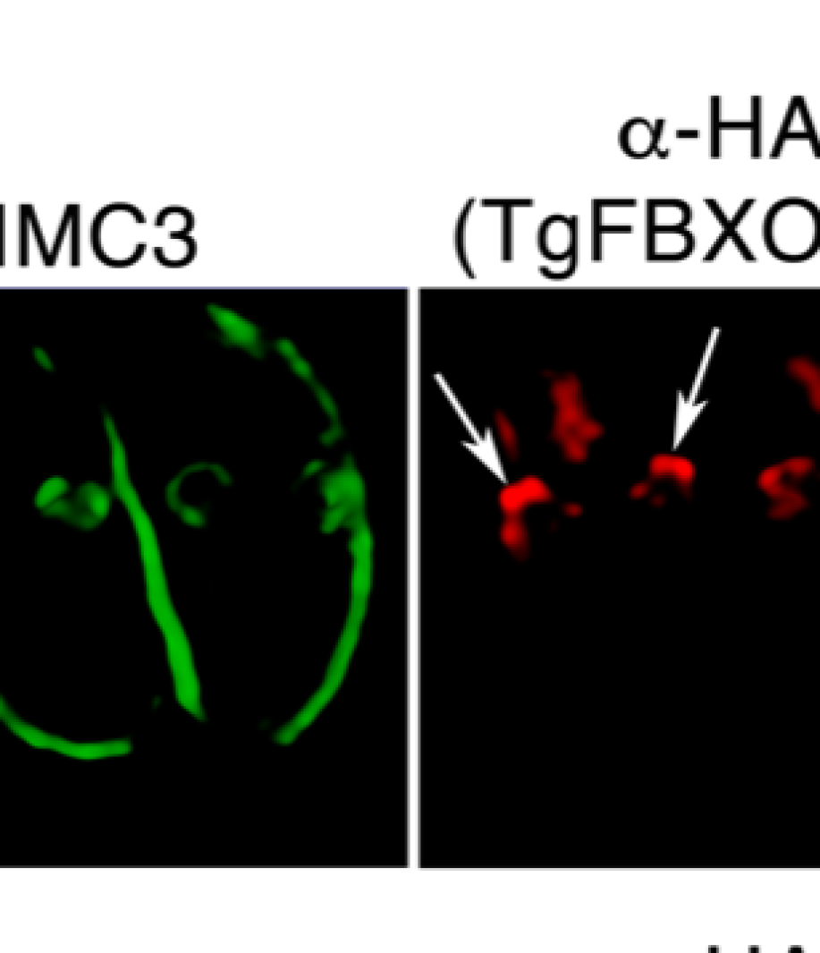
This figure is from Ira Blader’s lab; it shows the 3D arrangement of proteins and membranes of Toxoplasma gondii parasites undergoing replication. This image was acquired on the Leica SP8 STED super-resolution confocal microscope.
Panels A through C compare the three-dimensional locations of 3 different components of the parasite (IMC3, ISP1 and a-myc) to the location of the FBOX01 protein. The volume rendered image shows that FBOX01 protein is an initiation site for these other components. F-box proteins are known to control cell cycle and membrane trafficking and thus are important in the replication of this parasite which is the cause of the disease, toxoplasmosis.
Upcoming Events
No events scheduled.