Research
General research interests of the core faculty include all aspects of medical, radiological, and health physics.
Work within the diagnostic/interventional imaging group at the Canon Stroke and Vascular Research Center (CSVRC) emphasizes the development of new imaging systems and computerized instrumentation, as well as improvements in image processing. A variety of applications within these areas are currently being investigated.
Diagnostic Imaging
Projects which have been underway include the exploration of CMOS-based high resolution fluoroscopic detectors, application of new detector technology to cone-beam computed tomography (CBCT), development of new methods for evaluation of imaging systems using generalized cascade linear systems modeling, development of CBCT mathematical algorithms for region-of-interest CT, development of rapid, multiple scanning-beam techniques for improving radiographic contrast, the study of image magnification in fluoroscopy, the development of methods for quality control in diagnostic radiology, and the measurement, monitoring and reduction of patient dose during radiographic/fluoroscopic procedures.
MRI
Research in MRI centers on the high field facility (4.7 T) at Roswell Park where research into new contrast methods for evaluating cancer therapies and new MRI pulse sequences for improved imaging are being developed. A 3T MRI system dedicated to research will be available at the CTRC for collaborative translational projects.
Nuclear Medicine
Research by the nuclear medicine physics group has centered on new developments in PET imaging including new, higher resolution detection methods and improvements in imaging software and analysis. Collaborative work with the micro-PET scanner is also encouraged and multi-modality research is encouraged.
Radiation Therapy
The therapeutic physics group at Roswell Park Comprehensive Cancer Center has been doing research in a variety of areas related to IMRT and calculation and calibration of radiation dose distributions. Many of the projects are closely related to the busy clinical radiation oncology service provided at Roswell Park.
Solid State X-Ray Image Intensifier
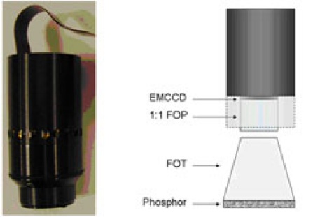

Currently the CSVRC is developing a Solid State X-ray Image Intensifier (SSXII) prototype that utilizes EMCCD (Electron Multiplying Charged Coupled Device) camera technology as well as interchangeable fiber optic tapers (FOT) to improve resolution in medical imaging. The interchangeable components allow for specific task-based optimization of the detector system, i.e. angiographic images and real-time fluoroscopic imaging, ROI cone beam computed tomography, or mammographic tomosynthesis. These new dynamic X-ray imagers have the potential to provide significant improvements over current state of the art detectors.
The SSXII can provide higher resolution angiographic images and lower noise fluoroscopic imaging compare to both flat panel detectors and standard X-ray image intensifiers.
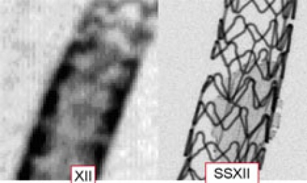

Two comparative images taken with a state-of-the-art XII and the new SSXII. Images are of an asymmetric stent, which is a new patient-specific device being developed by at the CSVRC for treatment of neurovascular aneurysms. Both images were acquired using the same exposure conditions and imaging geometry. The SSXII is shown to provide a potentially huge improvement over current technologies.
Patient-Specific Aneurysm Models
Another current area of interest for the CSVRC is in providing patient-specific aneurysm phantoms for investigating new minimally invasive endovascular image-guided interventional (EIGI) devices and evaluating the resulting flow modifications. The aneurysm phantoms are created during a multi-step process using segmented CT-scan data from a patient-specific aneurysm to produce a clear elastomer cavity with precisely the same dimensions. Such models allow for the implementation and evaluation of various EIGIs, such as stents, without the necessity of invasive surgery. Modifications to blood flow within the aneurysm are monitored and effective means of treatments are determined while greatly reducing risk to the patient.


Images of patient-specific aneurysm model creation. Left, Scale of the aneurysm form. Middle, Aneurysm model of silicon elastomer results after wax (red form) is liquified. Right, Fluoroscopic image of the contrast-filled model.
Other Research
We are developing a variety of real-time digital techniques for improved imaging in IGI radiographic procedures at reduced integral radiation doses. The projects combine the applied imaging physics of radiation sources and detectors with systems development of mechanical, electronic, optical, as well as computer control and image-processing capabilities.

A micro-angiographic projection image of stent-mesh in an aneurysm phantom.
Our group was the first to demonstrate clinical angiography with high resolution region of interest micro-angiographic detectors that we have developed. In collaboration with Canon, we were the first to demonstrate high resolution angiography on a direct conversion flat panel detector containing an amorphous selenium photoconductor rather than a phosphor detection layer.
We also developed imaging methods for investigating hemodynamic flow during catheter based procedures and we have worked on implementing patient-specific blood flow modification with specially developed stents designed to treat brain aneurysms. With Cone-beam Computed Tomography (CBCT), we provided a fully three-dimensional computer model of such an aneurysm and simulated a treatment plan using Computed Fluid Dynamic (CFD) analysis followed by the design, creation, and deployment of an actual patient specific stent.
Examples of other projects have included the exploration of light amplifiers for high resolution fluoroscopic detectors, application of new detector technology to CBCT mammography, development of new methods to evaluation imaging systems using generalized cascade linear systems modeling, development of CBCT mathematical algorithms for region-of-interest CT, development of rapid, multiple scanning-beam techniques for greatly improving radiographic contrast, the study of image magnification in fluoroscopy, the study of x-ray image intensifier distortion characteristics, the development of methods for quality control in diagnostic radiology, and the measurement and reduction of patient dose during radiographic procedures.
The therapeutic physics group based at Roswell Park Comprehensive Cancer Center has been doing research in a variety of areas related to intensity modulated radiation therapy (IMRT) and calculation and calibration of radiation dose distributions. Many of the projects are closely related to the busy clinical radiation oncology service provided at Roswell Park.
Research by the nuclear medicine physics group has centered on new developments in PET imaging including new, higher resolution detection methods and improvements in imaging software and analysis. Collaborative work with the micro-PET scanner and multi-modality research is encouraged.
Research in MRI is based at the high field facility at Roswell Park where research into new contrast methods for evaluating new cancer therapies and new MRI pulse sequences for improved imaging are being developed.